
Science | November 2, 2026 | Peer-reviewed preclinical research
Two kinds of ultrasound tell a more complete story
Researchers have reported a new approach to predicting how tumors respond to treatment by combining two different ultrasound measurements with deep learning. The study, published November 1 in the journal npj Imaging, examined images collected before chemo-immunotherapy in mouse models of breast cancer, sarcoma and melanoma. One ultrasound technique measured tissue stiffness; the other measured blood perfusion. The combined model reached an overall test accuracy of 0.930 in the research dataset, compared with 0.877 for the stiffness-based model and 0.825 for the perfusion-based model. These figures are promising but must be read in context. They describe a preclinical experiment, not a diagnostic service proven in human patients. The central scientific idea is that a tumor’s physical environment may reveal useful clues about how effectively treatment can reach and affect it.
Why tumor stiffness is biologically relevant
Cancer is not merely a collection of abnormal cells. A solid tumor exists within a surrounding environment of connective tissue, blood vessels, immune cells and other structures that can influence treatment. As tumors become mechanically stiff, pressure inside the tissue can compress blood vessels. That may reduce circulation, limit oxygen delivery and make it harder for some medicines to penetrate the affected area. Ultrasound shear wave elastography, abbreviated SWE, estimates tissue stiffness by measuring the speed at which mechanically generated shear waves move through it. This provides information about the physical properties of the tissue without requiring a surgical sample for that measurement. Yet stiffness alone cannot capture every aspect of tumor biology. Two lesions with similar mechanical characteristics may have different blood supplies or immune environments, which is why the researchers investigated a second imaging signal.
The second signal measures blood flow
Contrast-enhanced ultrasound, known as CEUS, uses microbubble contrast agents to visualize and quantify blood flow in tissue. In oncology research, this can provide a picture of perfusion, meaning how effectively blood reaches different parts of a tumor. Perfusion matters because circulating blood delivers oxygen, nutrients and many systemically administered drugs. Poorly perfused regions may be difficult to treat, but blood flow is only one part of a complex biological process. The study’s design treated perfusion and stiffness as complementary rather than competing indicators. The researchers reasoned that one measurement could reveal something the other missed. A stiff tumor may have impaired circulation, but the relationship varies across tumor types and conditions. By analyzing both image sets together, the model could potentially capture patterns associated with treatment response that would be less visible in either technique on its own.
How the models were constructed
The team first trained a convolutional neural network called CNN-ultra separately on shear wave elastography and contrast-enhanced ultrasound images. These networks were designed to learn visual patterns linked to subsequent treatment outcomes. The researchers then developed CNN-combi, which integrates information from both modalities. The published dataset contained 587 ultrasound images acquired before therapy, with responses categorized as responsive, stable or non-responsive. The reported counts were 175 responsive cases, 136 stable cases and 276 non-responsive cases. That imbalance matters because a model can appear accurate while performing unevenly across categories. The authors therefore reported class-specific results as well as overall accuracy. Importantly, the images came from experimental mouse models. They should not be described as hundreds of independent human patients, and image counts are not automatically equivalent to the number of biologically independent animals.
What the accuracy figures show and what they do not
The integrated model achieved a reported overall test accuracy of 93.0 percent. The authors reported class-specific accuracies of 98.3 percent for responsive tumors, 93.0 percent for stable tumors and 94.7 percent for non-responsive tumors. These results support the study’s central finding that the combination of stiffness and perfusion measurements improved predictive performance within the experimental framework. They do not establish that the same model would perform equally well in hospitals. Accuracy depends on the composition of the test data, imaging protocols, disease mix and how outcomes are defined. Performance can change when images come from different scanners, institutions or patient populations. Clinical usefulness also requires considering the consequences of errors. A mistaken prediction that a tumor will respond could be particularly consequential if it influenced treatment decisions. The study provides evidence of feasibility, not authorization to guide care.
Why treatment-response prediction is difficult
Oncologists routinely face uncertainty about whether a particular therapy will work for a particular tumor. Tumors vary genetically and structurally, and their response can change as treatment proceeds. A technique that identifies likely non-response before therapy begins could eventually help researchers design more efficient treatment strategies or identify patients who need closer monitoring. But predicting response is not the same as choosing the best treatment. A clinically useful system would have to demonstrate that its predictions improve decisions and outcomes compared with existing assessments. It would also need to account for the many variables missing from a pair of imaging measurements, including molecular biomarkers, patient health, treatment dose and prior therapies. The new research is best understood as an effort to add physical and vascular information to the broader precision-oncology toolkit.
What makes the combination technically interesting
The study illustrates a broader shift in medical artificial intelligence from relying on a single image type toward integrating multiple signals. Different measurement technologies reveal different aspects of the same biological object. Stiffness maps can suggest mechanical resistance, while contrast-enhanced scans can reveal vascular function. A neural network may learn interactions between those signals that are difficult to express as simple rules. The benefit is not automatic, however. Combining modalities increases the demands on data quality, alignment and model evaluation. If one image is noisy or collected under a different protocol, the system may learn misleading correlations. The research therefore raises practical questions about repeatability and standardization. Before clinical deployment, investigators would need to establish that the apparent improvement is robust rather than dependent on a particular experimental dataset.
The limits of mouse-model evidence
Mouse models are indispensable for exploring mechanisms and testing new ideas, but they do not reproduce the full diversity of human cancer. Tumor size, growth rate, immune environment and the way treatment is administered can differ substantially between laboratory animals and patients. The study also examined three experimental cancer categories rather than the broad range of clinical diseases that a general-purpose oncology tool might encounter. Those limits do not diminish the scientific result; they define its proper scope. A reasonable next stage would involve larger and more diverse datasets, independent testing, standardized ultrasound acquisition and prospective human studies where appropriate. Researchers would need to assess whether the model adds value beyond established clinical measures. Until then, no patient should interpret the reported 93 percent figure as a personalized probability that their own treatment will succeed.
How clinicians might eventually use such information
If later studies validate the approach, combined ultrasound measurements might help clinicians characterize tumors before treatment or monitor changes during a course of therapy. Ultrasound is attractive because it is widely used, can provide real-time information and avoids ionizing radiation. Contrast-enhanced techniques and elastography nevertheless require trained operators, appropriate equipment and clinical protocols. An AI model would also need transparent quality controls, clear intended uses and procedures for handling uncertain predictions. It should support clinical judgment rather than replace it. The most valuable application might not be a simple yes-or-no answer but a better understanding of which tumor environments create barriers to treatment. That knowledge could inform future studies of therapies designed to improve perfusion or modify mechanical conditions inside tumors.
The real advance is a research direction, not a cure
The November 1 publication offers a credible demonstration that two ultrasound-based biomarkers can be more informative together than separately in a preclinical setting. It does not report a new cancer cure, a successful human trial or a ready-to-deploy hospital product. Those distinctions are essential because artificial intelligence results can attract attention through a single impressive percentage. The study’s more durable contribution is methodological: it shows how physical measurements of a tumor and its blood supply can be integrated to investigate response to chemo-immunotherapy. Independent validation, human data and clinical outcome studies will determine whether the approach becomes useful beyond the laboratory. For now, the finding strengthens the case for studying the tumor microenvironment as a source of predictive information while leaving the crucial clinical questions open.
Frequently asked questions
Was the ultrasound AI tested on human cancer patients?
No. The reported study used preclinical mouse models of breast cancer, sarcoma and melanoma.
What accuracy did the combined model achieve?
The journal reports an overall test accuracy of 0.930, or 93.0 percent, within the research dataset.
Can this test now determine which cancer treatment someone should receive?
No. Clinical validation and assessment of patient outcomes are needed before such a model could guide treatment decisions.
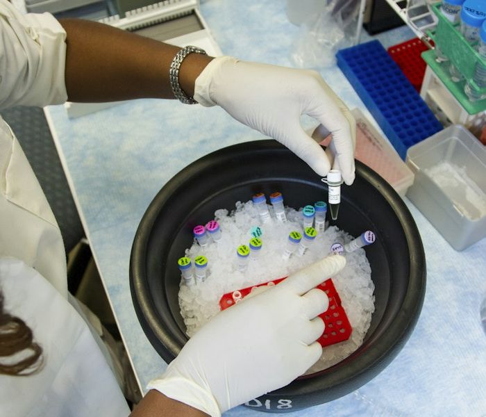
Primary source: Dimou and colleagues, npj Imaging, published November 1, 2026. Featured photograph: Elen Sher / Unsplash, illustrative clinical equipment.